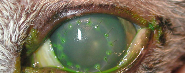
The recipient at a check-up 2 weeks following surgery. Fluorescein dye has been placed in the eye to visualize the intactness of the corneal epithelium.
The recipient at a check-up 2 weeks following surgery. Fluorescein dye has been placed in the eye to visualize the intactness of the corneal epithelium.
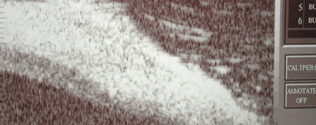
A sample ultrasound picture of a sequestrum. The white hyperechoic area shows exactly the thickness and location of the sequestrum within the cornea. This allows for a more accurate excision of the lesion to the exact depth needed.
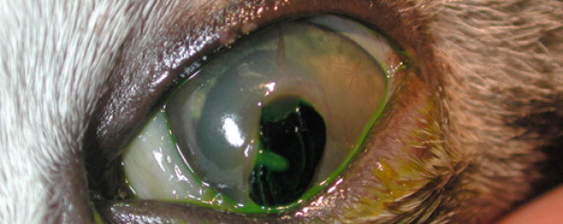
A patient with a very large sequestrum. It is the large dark kidney bean-shaped area in the lower right hand portion of the cornea.
Musculoskeletal soft tissue grafts (tendons and fascia) are available: including patella (bone-tendon-bone) grafts, superficial digital flexor tendon grafts and the common calcaneal tendon w/ bone block (Achilles’ tendon) graft.
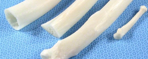
Shafts and whole bones are tissues that are generally not possible to obtain as autografts. They are most commonly used for replacement of large bone sections affected by osteosarcoma in an effort to avoid amputation (limb-sparing procedure).
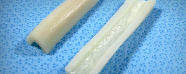
Cortical Struts are longitudinal sections of cortical bone from canine tibia, radius, or femur. They will have a flat to semi-round cross-section. The thickness of the cortex will be at least 3mm. They are either ~0.5 or 1.0cm wide and ~3.0 – 5.0cm in length.
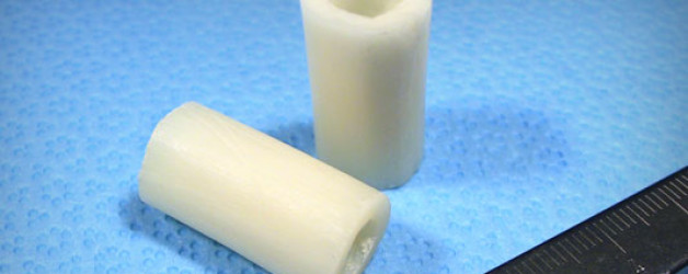
Cortical Segments are femur, tibia or radius ring sections.
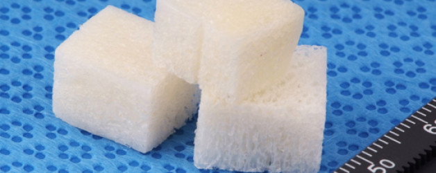
Cancellous Blocks are made from extremely dense cancellous bone typically found in the condyles and plateaus of femur and tibia.
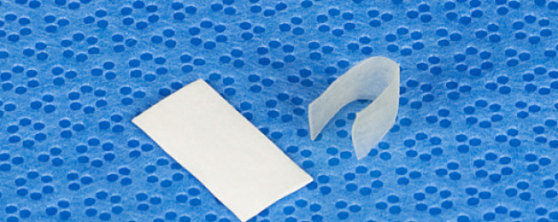
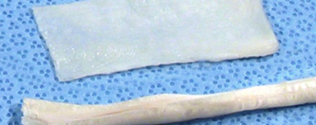
Ossiflex™ Bone Membranes shown freeze-dried (left) and rehydrated and bent (right).
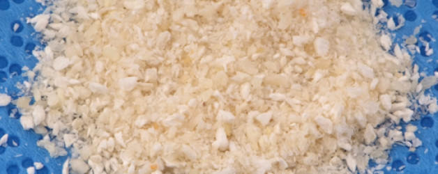
Demineralized Bone Matrix (DBM) is osteoinductive. It is made from cortical bone that has been ground to particle sizes of 1.25 mm or less. The mineral content is reduced using hydrochloric acid. This leaves the matrix and the acid-resistant, endogenous bone growth factors in place (available as frozen).