Images courtesy of Dr. Jason Bleedorn, MS, DVM, DACVS of the School of Veterinary Medicine at the University of Wisconsin – Madison and Dr. Allen Johnson, DVM, DACVS of Animal Surgical Clinic in Seattle, WA.
Using bone graft in osteotomy sites is quick, easy, and the best way to speed healing in orthopaedic surgeries. In most surgical and referral practices, treatment of canine cranial cruciate ligament disease constitutes a large portion of the caseload. Ensuring that these patients recover quickly and without post-operative complications will enhance a surgeon’s reputation and undoubtedly grow their practice. VTS can help!
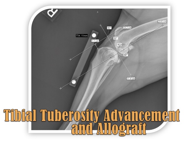
Tibial tuberosity advancement is becoming a popular choice among orthopaedic surgeons as a treatment for canine cruciate ligament disease. TTAs are a perfect surgery for using allograft, as the procedure leaves a large osteotomy site (in a wedge shape). In one published study, 84% of TTA cases using allograft healed by 12 weeks (with an average healing time of 9.4 weeks)1. In a separate study of TTAs performed without allografts, only 59% of patients were healed by 14 weeks2. Below, see a step-by-step guide for using VTS’ bone graft in a TTA procedure.
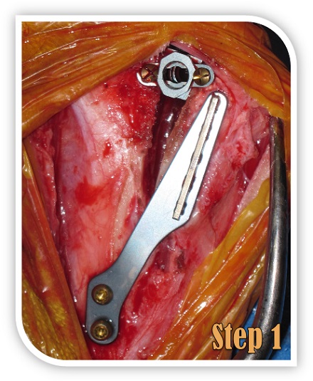
This photograph illustrates a nearly finished TTA surgery. The wedge osteotomy site is to the left, caudal to the TTA cage. Ensure the osteotomy site has been irrigated thoroughly.
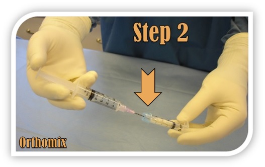
If you are using freeze-dried graft, rehydrate the graft while still in the syringe by injecting about 0.5 to 1cc of patient blood or sterile saline through the blue syringe cap. If frozen graft is used, rehydration is not necessary.

Express the graft from the syringe into the bleeding osteotomy site. An elevator or other instrument can be used to pack the graft, and graft rehydrated with patient blood will begin to clot, facilitating graft placement.

The graft should also be placed inside the TTA cage. VTS’ bone graft is both osteoconductive and osteoinductive. By placing the graft inside of the implant, bone can grow in and around the implant, strengthening the repair.

A fully grafted TTA site just prior to closure. The particulate has been packed into the osteotomy site, the implant cage, and no portion of the osteotomy site is left unfilled.
Whether you do a few TTA surgeries a day or only every few months, VTS’ allograft products can help you and your practice. VTS is proud to offer graft products that fit every budget – the possibilities are endless – and all of our products promise better surgical outcomes for your patients.
References (click for full-text PDF)
1Lafaver S, Miller NA, Stubbs WP, Taylor RA, Boudrieau RJ. Tibial tuberosity advancement for stabilization of the canine cranial cruciate ligament-deficient stifle joint: Surgical technique, early results, and complications in 101 dogs. Veterinary Surgery. 36:573-586, 2007.
For further reading, please visit our references page!