Maxillary canines can leave large bone voids when extracted. The loss of this tooth and surrounding bony structures can lead to maxillofacial changes, including “lip catch.” Once healed, mandibular canines can cause trauma to the tissue covering the extraction site or upper lip. The use of bone graft to fill the extraction site can prevent these changes in the maxilla.
The most effective way to fill such a large extraction site is to use a barrier (Ossiflex or Fascia) and particulate bone graft (Periomix).
1. To rebuild the convexity of the maxilla following a maxillary canine extraction, fill the empty alveolus with Periomix. The rebuilding of convexity helps the mandibular canine to fit without catching the lip or traumatizing the surrounding tissues or other physiological structures.
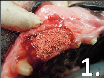
Periomix in a maxillary canine extraction site. Rehydrate the freeze-dried bone particles with a few drops of patient blood and pack into the site.
2. VTS’ flexible, natural bone membrane, Ossiflex, or pieces of Fascia can be placed on top of the Periomix to prevent the particulate granules from dispersing and facilitate bone healing under the protection of a barrier. This is known as Guided Bone Regeneration.
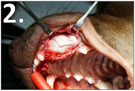
Fascia pieces cover the Periomix in a maxillary canine extraction site. Rehydrate the freeze-dried fascia or membrane with sterile saline before placing into the site.
3. Once the particulate and soft tissue or membrane are in place, close the flap created for the extraction. Then, proceed to full closure.
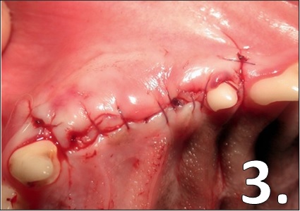
Maxillary canine extraction site at full closure. Note that the maxillary convexity created by the process of Guided Bone Regeneration is visible once closure is complete.
Images 1 and 3 courtesy of Dr. Tony Caiafa, DVM, DDS, DAVDC and Dr. Loïc Legendre, DVM, DAVDC, DEVDC. Image 2 courtesy of Dr. Rocco Mele, DVM. canlı bahis