This case courtesy of Dr. Rocco Mele, DVM of Eastpoint Pet Clinic in Tucson, Arizona
Oronasal fistulas (ONFs), a communication between the oral cavity and the nasal passages, are a common malady seen by both veterinary dental specialists and general practice veterinarians. Oronasal fistulas can be caused by trauma (such as a bite wound), or sometimes by extraction of large teeth; or by bone loss secondary to periodontal disease.
Riley was referred to Eastpoint Pet Clinic with an ONF (palatal and apical to tooth #104). This fistula occurred secondary to the surgical extraction of her maxillary canine tooth by a referring veterinarian several weeks earlier.
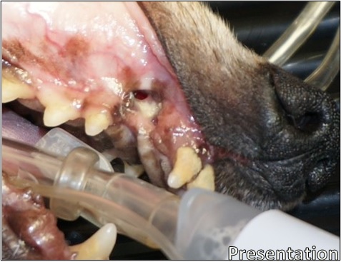
Oronasal fistulas can be painful and uncomfortable to pets. Bits of food can become lodged in the defect or enter the nasal passages, causing infection and discomfort. Riley was likely dealing with an infection when this photograph was taken; purulent discharge can be seen in and around the defect.
Large tooth extractions with oronasal fistulas like this one can result in significant bone defects in the maxilla, providing a passageway for food (and infection) to enter the nasal cavity, and poor Riley was no exception.
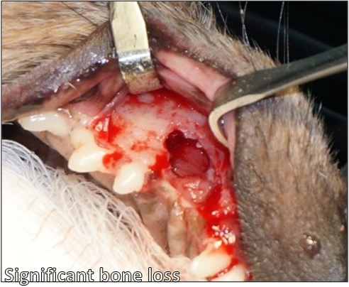
Riley’s oronasal fistula, with soft tissues fully retracted. Here, the mouth’s communication with the caudal nasal passages can be fully appreciated. With a bony void of this size, failure to fill the site with bone graft can result in soft tissue collapse, surgery site dehiscence, and necessitate further surgery.
The repair of oronasal fistulas is perfect for use of VTS’ Ossiflex, a thin, flexible bone membrane. Riley’s defect was repaired using an Ossiflex on the right side, which was trimmed to fit the defect after it was rehydrated with sterile saline.
The rehydrated membrane is placed over the defect and sutured in place. The membrane provides a rigid barrier that allows for bone growth to happen in a protected environment. The membrane closes the abnormal opening between the oral and nasal cavities. It will slowly ossify, providing support for Riley’s soft and hard tissue to regenerate.
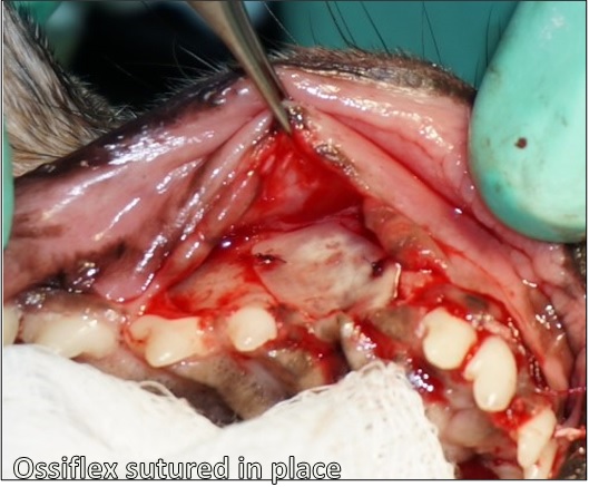
The trimmed Ossiflex is sutured in place using absorbable suture. Stabilization ensures the membrane will remain in place following flap apposition, as the surrounding tissue heals.
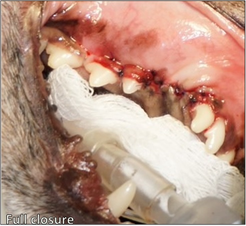
After the Ossiflex is secured and the defect is covered completely, the surrounding soft tissues can be replaced and sutured closed with a tension-free flap.
At seven months post-operative, Riley has healed beautifully. The soft tissue covering the defect site, per Dr. Mele, is solid and firm to the touch. Riley is able to eat, drink, and play as she did prior to developing the fistula with no risk of food entering her nasal passages.

Riley has been anesthetized for recheck radiographs at 7 months post-operative. The area where the defect was has completely healed, and the soft tissue appears healthy and strong, and maintains gingival margins similar to that of her surrounding teeth, preventing traumatic injury from her lower canines.
Radiographically, ONFs can be difficult to visualize. Below, Riley’s pre-operative and 7 month post-operative films illustrate tremendous bone growth. A “bridge” of new bone can be seen across the space previously occupied by her maxillary canine. As Riley continues to heal, that bridge will likely continue to grow, strengthening her maxillary bone structure.

Radiographs of pre-operative and 7 months post-operative. The circle highlights the bridge of new bone that has grown over the void left by the fistula and maxillary canine extraction.
Ossiflex is ready to cover your largest bony defects! Available in five sizes; thin enough to cut and suture, yet strong enough to resist food pressure and provide an immobile substrate for bone regeneration. Our flexible bone membrane is the answer for any patient with an ONF!