Case courtesy of Dr. Matt Corse, DVM, MS, ACVS and Dr. Kenneth Greenwood, DVM, MS, ACVS of Northlake Veterinary Surgery, Clarkston, GA
Surgical revisions are a reality of veterinary orthopaedics. Patients are often referred to a specialist when something goes awry. In a referral practice, patients often present needing a second, third, or even fourth surgical procedure on the same site. This is especially common with orthopaedic trauma cases; complex fractures can result in nonunion or necessitate additional surgical interventions to ensure fixation.
This was the case for “Razor,” a 14 month-old Laborador who was referred to Northlake Veterinary Surgery. Razor presented with an external fixator in place due to a fracture repair that had failed. Radiographs were first taken with the external fixator in place.
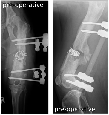
Cranial caudal and lateral views of the fracture site. Circlage wire was placed in an earlier revision by the surgeon who had performed the initial repair. In the lateral view, bone can be seen attempting to grow around the failing implant. This causes non-union and instability. In this case, it has also resulted in re-fracture, collapse, and shortening of the femur.
The external fixator was then removed, and additional views were taken.

The re-fracture is more obvious with the external fixator removed. Despite bone attempting to grow around the implant, union has not been achieved and the repair remains highly unstable.
A surgical femur fracture revision was performed using allografts from Veterinary Transplant Services. Surgeon Dr. Matt Corse used a canine metatarsal segment to bridge the defect left behind once the surgical site was fully debrided. The site was then packed with Orthomix and closed.

Radiographs taken immediately post-operative show the segmental allograft in place (seen near the center), secured to the plate with the two center bone screws. The particulate fragments seen around the segment is VTS’ osteoinductive particulate bone graft, Orthomix.
The segmental and particulate allografts are both osteoinductive and osteoconductive, and facilitate remarkable bone healing. Razor recovered well and eventually the fracture site had healed enough to have the plate removed. Union was acheived at the fracture site, and the repair stable. In these follow-up radiographs, the fracture site is hardly visible.
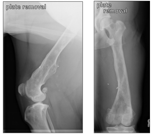
At approximately one year post-operative, the fracture site is completely healed. The repair is stable enough for the plate to be removed. New bone has grown in and around the fracture site and union has been achieved. The patient’s limb has been restored to its normal length and is able to fully support the weight of the dog.
Have a difficult non-union or surgical revision looming on your surgical calendar? Call VTS today and order bone graft products to ensure your patients have an optimized chance to heal quickly and completely.